Meat is a widely consumed product worldwide and to meet this demand, new tools and technologies are under development for more efficient and effective measurement and quality control. Guaranteeing tenderness would increase consumers’ confidence and significantly influences the decision to return. Several studies have shown that consumers would be ready to pay more for a steak with higher tenderness and quality. Due to this, the beef industry is giving increasing importance to quality assurance and analytical techniques that allow product quality control.
In France, the Blaise Pascal University and INRA developed in 2010 an image processing method to predict beef tenderness, collagen and lipids contents in the meat, using images of semimembranosus muscle slices acquired under visible and ultraviolet lighting. A statistical technique was implemented as a method to relate the distribution of intramuscular connective tissue (which is one of the major contributors to the toughness of beef), characterized by image analysis, to sensory tenderness, collagen and total lipids contents. For that, two groups of Salers and Holstein cull cows were fattened on the same diet and slaughtered at the same body fatness condition scored by palpation. Samples of semimembranosus muscle were taken for:
- collagen and total lipid determinations: assessed chemically.
- tenderness evaluation: assessed by a panel of 12 trained persons to evaluate during five sessions the tenderness, juiciness and flavour of the meat.
- Image analysis: muscle sample of each animal was cut into a cube and placed in a methacrylate box, which is shown in the Figure 1A.
The images of the meat cubes surfaces were made using a photographic measurement bench equipped with a camera, two white light lamps and an ultraviolet lamp (Figure 1B). The white light lamps were equipped with a polarising filter in order to reduce the reflectance of the sample. The ultraviolet lighting allowed to highlight the connective tissue network on a wavelength of 380 nm, which was identified by fluorescence spectroscopy as the longest excitation wavelength for fluorescence of collagen. A total of 96 images (24 samples × 2 faces of each cube × 2 light types) were captured and processed for the extraction of the image parameters.
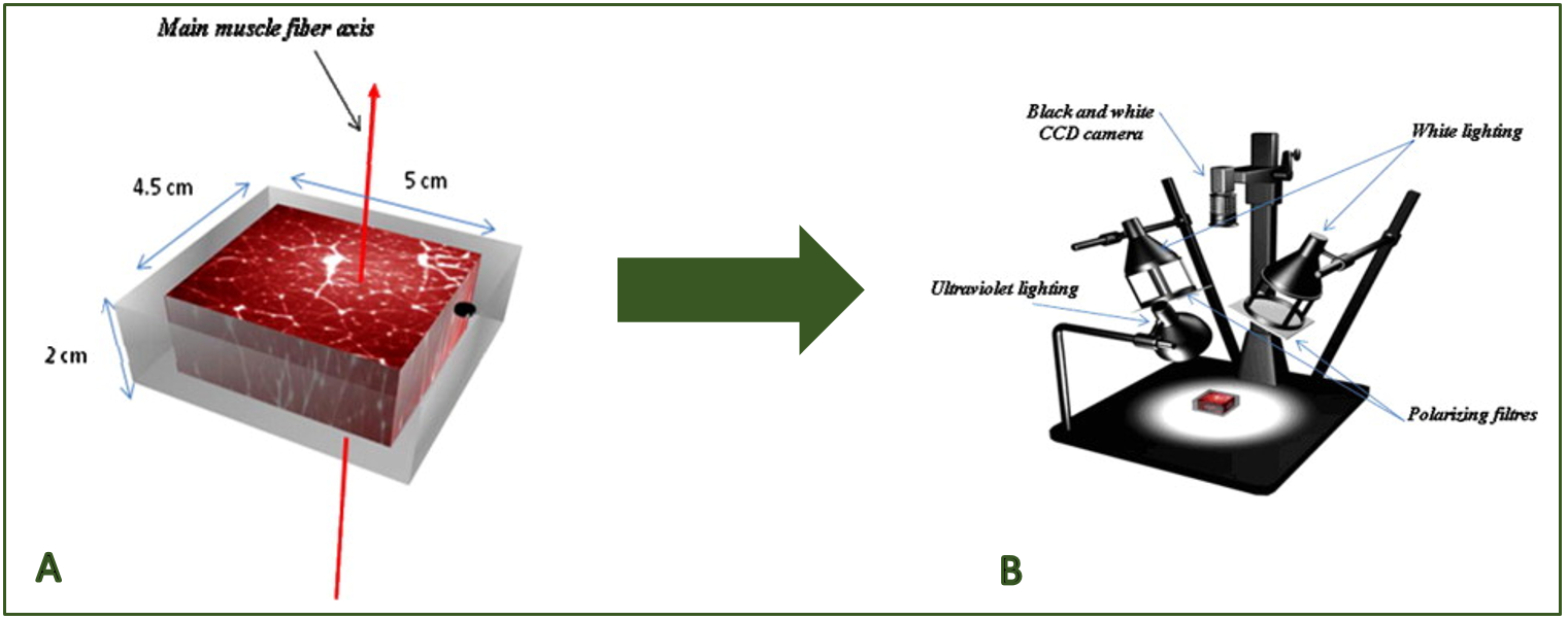 Figure 1: A) muscle sample preparation. B) photographic measurement bench (Adapted from El Jabri et al., 2010).
The results showed that the intramuscular connective tissue image parameters were found to be good predictors of beef tenderness (R2 = 0.89), collagen (R2 = 0.82) and lipids contents (R2 = 0.91). The image analysis method can be regarded a promising technique for prediction of meat tenderness, as the tenderness evaluation by sensory analysis with trained panelists can be subjective, expensive and time consuming, making it impossible to use as a routine analysis in the meat processing industry. However, further research is needed to establish a more reliable prediction of meat tenderness through image analysis.
*Post suggested by Alessandro Ferragina (Teagasc, Ireland).
Authors: Jakeline Vieira Romero, Virginia C. Resconi, Alessandro Ferragina.
Impact on:
 | Image analysis method could be more accessible and cheaper for the meat industry. |
 | |
 | Less subjective and more accurate information on meat tenderness. |
 |
Source of information:
El Jabri, M. et al. (2010). Journal of Food Engineering. 96:316-322. https://doi.org/10.1016/j.jfoodeng.2009.08.006